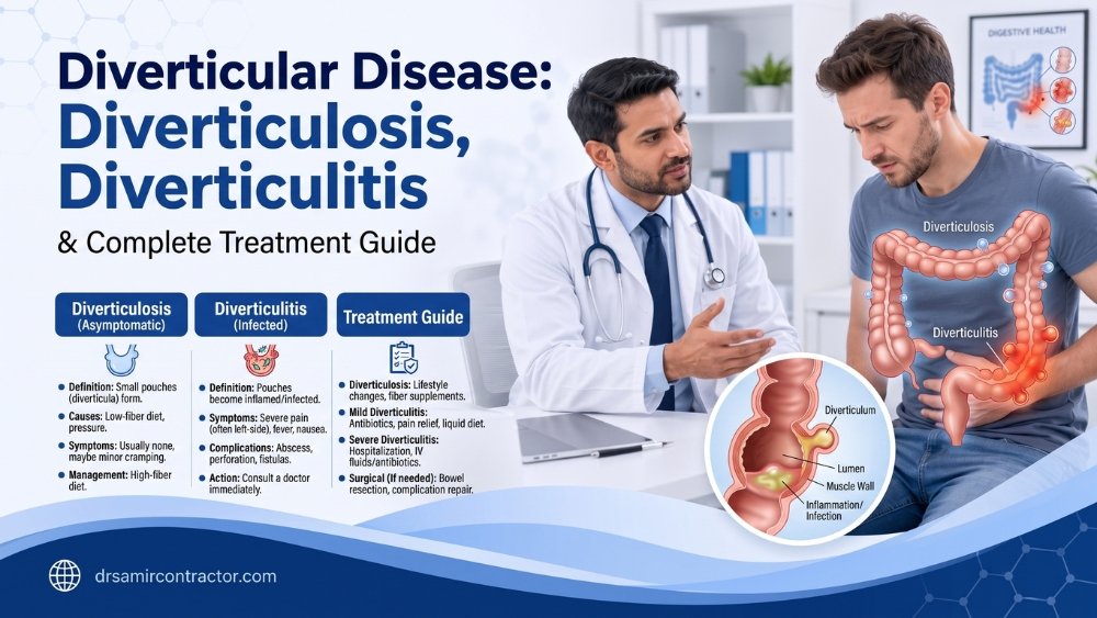
Diverticular disease refers to the spectrum of conditions that arise when small pouches - called diverticula - form in the wall of the colon. When the pouches sit quietly without causing trouble, we call it diverticulosis. When they become inflamed or infected, it becomes diverticulitis - a condition that can range from mild left-sided pain managed at home to a surgical emergency involving perforation or abscess. Once considered a disease of Western countries, diverticular disease is now rising sharply in India as diets shift away from traditional high-fibre foods. This page covers the full spectrum: what causes colon diverticula, how to recognise diverticular disease symptoms, when you can manage conservatively, and when surgery becomes necessary.
✦ Quick Answers
Understanding the Spectrum: From Diverticulosis to Complicated Diverticulitis
Diverticular disease is not a single diagnosis - it is a spectrum. Understanding where you fall on that spectrum determines everything about your treatment. The vast majority of people with colon diverticula never know they have them. A smaller proportion develop symptoms. And a still smaller group faces complications that require urgent intervention. Here is how the spectrum breaks down:
- Diverticulosis (asymptomatic) - Diverticula are present but cause no symptoms. This is an incidental finding on colonoscopy or CT scan. No treatment is needed beyond dietary optimisation to prevent progression.
- Symptomatic uncomplicated diverticular disease (SUDD) - Diverticula cause chronic or recurrent left lower abdominal pain, bloating, and altered bowel habits without overt inflammation. This overlaps considerably with irritable bowel syndrome, making diagnosis challenging.
- Acute uncomplicated diverticulitis - A diverticulum becomes inflamed or micro-perforated, causing localised pain, fever, and elevated inflammatory markers. CT scan shows pericolic fat stranding without abscess or free perforation.
- Acute complicated diverticulitis - Inflammation progresses to abscess formation, frank perforation with peritonitis, fistula (abnormal connection to bladder, vagina, or skin), or stricture causing bowel obstruction. This is the stage that often requires surgery.
What Causes Diverticula to Form?
Diverticula develop at points of weakness in the colon wall - specifically where small blood vessels (vasa recta) penetrate the muscular layer to supply the mucosa. When intraluminal pressure is high (typically from straining against hard, low-bulk stool), the mucosa herniates through these weak points, forming a pouch. This is why the sigmoid colon - the narrowest segment with the highest pressures - is the most commonly affected site.
Several factors contribute to diverticula formation:
- Low-fibre diet - This is the single most important modifiable risk factor. A low-fibre diet produces small-volume, hard stool that requires higher colonic pressures to propel forward, directly increasing the mechanical stress on the colon wall.
- Age-related collagen changes - With ageing, colonic wall elasticity decreases and the muscular layer thickens, creating higher resting pressures and more susceptible weak points.
- Obesity - Increased intra-abdominal pressure and chronic low-grade inflammation associated with visceral fat raise the risk of both diverticula formation and diverticulitis.
- Sedentary lifestyle - Physical inactivity reduces colonic motility and is independently associated with a higher rate of diverticular complications.
- Smoking - Impairs colonic wall integrity and microcirculation, increasing the risk of perforation.
- Medications - Regular use of NSAIDs (ibuprofen, diclofenac), corticosteroids, and opioids is associated with increased diverticulitis risk and a higher rate of complicated episodes.
Diverticulosis vs Diverticulitis - Key Differences
Patients frequently confuse these two terms. The distinction is clinically critical because management differs entirely.
| Feature | Diverticulosis | Diverticulitis |
|---|---|---|
| Definition | Diverticula present without inflammation | Diverticula with active inflammation or infection |
| Symptoms | Usually none; occasional mild bloating | Severe left lower abdominal pain, fever, nausea |
| Fever | Absent | Often present (38°C or higher) |
| Blood tests | Normal CRP and white cell count | Elevated CRP and white cell count |
| CT scan findings | Diverticula only, no fat stranding | Pericolic fat stranding, wall thickening, possibly abscess |
| Treatment | High-fibre diet, no medication needed | Antibiotics, bowel rest, possibly surgery |
| Urgency | Routine - dietary counselling | Urgent - same-day medical assessment needed |
| Complications | Risk of bleeding; low risk of progression | Abscess, perforation, fistula, stricture |
Diverticular Disease Symptoms - What to Watch For
The symptoms of diverticular disease vary significantly depending on where you fall on the spectrum. Recognising these patterns helps you determine whether self-management is appropriate or whether you need urgent evaluation.
Chronic Symptomatic Diverticular Disease
Patients with symptomatic uncomplicated diverticular disease describe a recurring pattern: left-sided lower abdominal cramping or aching that worsens after meals and improves after passing stool or gas. There is often associated bloating, a sense of urgency to pass stool, and alternating constipation and loose motions. The symptoms mimic IBS so closely that many patients carry an IBS diagnosis for years before diverticula are identified on colonoscopy or CT.
Acute Diverticulitis
An acute attack is unmistakable. The pain is constant (not cramping), localised to the left lower abdomen, and accompanied by fever, loss of appetite, and sometimes nausea or vomiting. The area is tender to touch. Some patients describe it as a “left-sided appendicitis.” In older or immunosuppressed patients, the presentation can be more subtle, with less fever and less localised pain, making clinical suspicion essential.
Diverticular Bleeding
Diverticular bleeding is the most common cause of significant lower gastrointestinal haemorrhage in adults over 60. It typically presents as sudden, painless, large-volume rectal bleeding - bright red or maroon blood. The bleeding occurs when a small artery (vas rectum) at the neck of a diverticulum erodes. While alarming, diverticular bleeding stops spontaneously in approximately 75-80% of cases. However, patients with significant blood loss, haemodynamic instability, or recurrent bleeding episodes require hospitalisation and may need colonoscopic intervention, angiographic embolisation, or surgery.
The Hinchey Classification - Grading Complicated Diverticulitis
When diverticulitis moves beyond simple inflammation, surgeons use the Hinchey classification to grade the severity of complications. This grading system directly determines the treatment approach.
| Hinchey Stage | Description | Typical Management |
|---|---|---|
| Stage I | Pericolic or mesenteric abscess (confined) | IV antibiotics; CT-guided percutaneous drainage if abscess > 3-4 cm |
| Stage II | Pelvic, distant, or retroperitoneal abscess | CT-guided drainage + IV antibiotics; elective surgery after resolution |
| Stage III | Purulent peritonitis (generalised pus without free faecal contamination) | Emergency surgery - resection with or without primary anastomosis; laparoscopic lavage in select cases |
| Stage IV | Faecal peritonitis (free perforation with stool in abdomen) | Emergency surgery - Hartmann’s procedure (resection with end colostomy) or resection with primary anastomosis in experienced centres |
Hinchey I and II are managed non-operatively in the majority of cases, with surgery reserved for failure of drainage or recurrence. Hinchey III and IV are surgical emergencies where the priority is source control - removing the diseased segment and managing peritoneal contamination.
Diverticular Disease vs IBS - The Overlap Problem
One of the most common diagnostic challenges is distinguishing symptomatic diverticular disease from irritable bowel syndrome. Both conditions produce left-sided abdominal pain, bloating, and altered bowel habits. Both are common in the same age group. And - importantly - both can coexist in the same patient. Here is how clinicians differentiate them:
- Structural vs functional - Diverticular disease involves visible structural pouches; IBS has no structural abnormality. CT or colonoscopy shows the difference.
- Inflammatory markers - During a diverticulitis flare, CRP and white cell count are elevated. In IBS, these markers remain normal even during symptom flares.
- Fever - Present in diverticulitis, absent in IBS.
- Age of onset - IBS typically begins before age 40; diverticular disease symptoms usually begin after 50, though younger-onset cases are increasing.
- Response to antibiotics - Diverticulitis improves with antibiotics; IBS does not.
When symptoms overlap and the diagnosis is unclear, a CT scan of the abdomen during a symptomatic episode is the most useful investigation. A colonoscopy should be performed 6-8 weeks after any acute episode resolves, both to confirm the extent of diverticular disease and to exclude underlying colorectal cancer or polyps.
Red Flags - Seek Urgent Medical Attention
If you have known diverticular disease or suspect diverticulitis, seek immediate evaluation if you experience:
- Severe, constant left lower abdominal pain with fever above 38°C
- Generalised abdominal pain with rigidity or guarding (abdomen hard and tender all over)
- Heavy rectal bleeding - passing large amounts of bright red or maroon blood
- Inability to pass stool or gas - possible bowel obstruction from stricture
- Passage of stool or gas through the vagina or urine - suggests a fistula
- Pneumaturia (air bubbles in urine) - a hallmark sign of colovesical fistula
- Rapid heart rate, dizziness, or fainting with rectal bleeding - signs of significant blood loss
- Worsening pain despite antibiotics after 48-72 hours of treatment
Reassuring Signs - Likely Manageable at Home
Your diverticular symptoms are more likely benign and manageable if:
- The pain is mild, intermittent, and relieved by passing stool or gas
- There is no fever or the fever is low-grade and resolving
- You can tolerate fluids and light food without vomiting
- There is no blood in the stool
- The pain pattern is familiar from previous episodes that resolved with conservative measures
- You have already been evaluated and have a confirmed diagnosis of uncomplicated diverticular disease
Even with reassuring features, if you have not been previously evaluated, a first episode of suspected diverticulitis warrants medical assessment to confirm the diagnosis and exclude other conditions.
Treatment of Diverticular Disease - The Full Approach
Conservative Management for Diverticulosis and SUDD
The cornerstone of managing diverticulosis and symptomatic uncomplicated diverticular disease is dietary and lifestyle modification:
- High-fibre diet - 25 to 30 grams of fibre daily from whole grains, vegetables, fruits, and legumes. Fibre increases stool bulk, reduces intraluminal pressure, and decreases the risk of diverticulitis by an estimated 40%.
- Adequate hydration - 2 to 3 litres of water daily. Fibre without adequate water can paradoxically worsen constipation.
- Regular physical activity - At least 30 minutes of moderate exercise daily. Physical activity independently reduces diverticulitis risk.
- Weight management - BMI above 30 is a significant risk factor for complicated diverticulitis.
- Avoid unnecessary NSAIDs - Regular NSAID use increases the risk of perforation. Use paracetamol as the first-line analgesic when possible.
A note on seeds and nuts: The longstanding advice to avoid seeds, nuts, popcorn, and corn has been conclusively debunked. Multiple large studies have shown no increased risk of diverticulitis from these foods. In fact, nut consumption may be protective. There is no dietary restriction on these items.
Medical Treatment for Acute Diverticulitis
Uncomplicated diverticulitis (Hinchey 0 or mild Hinchey I without significant abscess) in an otherwise healthy patient can often be managed with oral antibiotics (a combination covering gram-negative and anaerobic bacteria), clear liquid diet progressing to low-residue as pain improves, and rest. Patients who are immunosuppressed, elderly, unable to tolerate oral intake, or whose symptoms fail to improve within 48 hours require hospital admission for intravenous antibiotics and monitoring.
When Does Diverticular Disease Need Surgery?
Surgery becomes necessary in several scenarios:
- Emergency indications - Hinchey III or IV (peritonitis from perforation), uncontrolled diverticular bleeding, and complete bowel obstruction from stricture. These require urgent or emergency colorectal surgery.
- Failed non-operative management - Abscess that does not respond to percutaneous drainage and antibiotics, or persistent symptoms despite maximal medical therapy.
- Recurrent diverticulitis - After 2-3 confirmed episodes of acute diverticulitis, especially in younger patients (under 50) who face a lifetime of recurrence risk, elective sigmoid colectomy is recommended.
- Complications - Fistula (colovesical, colovaginal, colocutaneous), chronic stricture causing partial obstruction, or inability to exclude malignancy.
The preferred surgical approach is laparoscopic sigmoid colectomy - removal of the diseased sigmoid segment with primary anastomosis (reconnection of the bowel). In experienced hands, this can be performed with a hospital stay of 3-5 days and return to normal activity within 2-4 weeks. In emergency settings with heavy contamination (Hinchey IV), a Hartmann’s procedure - sigmoid resection with end colostomy and closure of the rectal stump - may be the safest option, with colostomy reversal planned 3-6 months later.
Dealing with Diverticular Disease?
Whether you need dietary guidance for diverticulosis, medical management of a diverticulitis episode, or a surgical opinion on recurrent attacks, Dr Samir Contractor provides comprehensive care at Sterling Hospital, Vadodara.
Diagnosis - How Is Diverticular Disease Investigated?
The diagnostic pathway depends on whether you are in the middle of an acute episode or being assessed between episodes:
During an Acute Episode
- Clinical assessment - History, abdominal examination, temperature, and vital signs.
- Blood tests - Full blood count (raised white cells), CRP (elevated), renal function, and lactate if perforation is suspected.
- CT scan of the abdomen and pelvis with contrast - The gold standard for diagnosing acute diverticulitis. It confirms the diagnosis, stages severity using the Hinchey classification, identifies abscess, and excludes other causes of acute abdominal pain.
Important: Colonoscopy should NOT be performed during an acute diverticulitis episode due to the risk of perforation. It is typically scheduled 6-8 weeks after recovery.
Interval Assessment (Between Episodes)
- Colonoscopy - Performed 6-8 weeks after an acute episode to assess the extent of diverticular disease, exclude coexisting polyps or cancer (which can mimic diverticulitis on CT), and plan long-term management.
- CT colonography - An alternative if colonoscopy is incomplete or the patient cannot tolerate endoscopy.
Diverticular Disease in India - A Rising Concern
Diverticular disease was historically considered rare in India and other Asian countries. That is no longer the case. Over the past two decades, gastroenterology centres across urban India have reported a significant increase in diverticular disease incidence, and the pattern differs from Western populations in important ways:
- Westernisation of diet is the primary driver - The traditional Indian diet - built on whole grains (rotla, bajra, jowar), dal, vegetables, and seasonal fruits - is inherently high in fibre (30-40 grams daily). As urban Indians increasingly consume maida-based foods (bread, biscuits, naan, pizza), processed snacks, refined rice, and less fibre-rich vegetables, the dietary fibre intake has dropped to 10-15 grams daily in many households. This mirrors the dietary pattern that drove the diverticular disease epidemic in Western countries decades ago.
- Right-sided diverticula are more common in India - While sigmoid diverticulitis predominates in Western populations, Indian and Asian patients have a higher proportion of right-sided (caecal and ascending colon) diverticula. Right-sided diverticulitis can mimic appendicitis, leading to diagnostic confusion.
- Younger age of presentation - Indian data shows a notable proportion of diverticular disease cases in patients under 50, likely related to the early adoption of low-fibre diets by younger urban populations.
- Under-diagnosis is common - Many patients with chronic left-sided abdominal pain and altered bowel habits are diagnosed as “IBS” or “acidity” without imaging. A CT scan or colonoscopy often reveals unsuspected diverticular disease.
- Limited awareness among patients - Most Indian patients have never heard of diverticular disease. Unlike conditions such as piles or gallstones, diverticular disease does not feature in common health awareness. Patients often present only when complications (abscess, bleeding, or perforation) have already developed.
High-Fibre Indian Diet for Diverticular Disease
The good news is that the traditional Indian diet - when followed properly - is one of the best diets in the world for diverticular disease prevention and management. Here are specific, practical dietary recommendations:
- Grains: Prefer rotla, bajra roti, jowar roti, nachni (ragi) roti, and brown rice over maida roti, white bread, or polished rice
- Dal and legumes: Moong, tuvar, masoor, chana, rajma, and sprouted pulses are excellent fibre sources - aim for one serving at every main meal
- Vegetables: Generous portions of leafy greens (palak, methi, tandaljo), gourd vegetables (dudhi, turai, karela), and root vegetables (suran, shakarkand)
- Fruits: Guava, papaya, apple (with skin), pear, banana, and seasonal fruits - 2-3 servings daily
- Seeds and supplements: Isabgol (psyllium husk) 1-2 teaspoons with water at bedtime; flaxseed (alsi) in chutneys; til (sesame) in laddoos
- Hydration: Buttermilk (chaas), nimbu pani, coconut water alongside 2-3 litres of plain water daily
Gujarati & Hinglish ma Puchtachha Sawalo
Diverticular disease ma aapna mota aantarda (colon) ni diwal ma nana nana khisaa (pouches) bani jaay chhe - aane diverticula kahe chhe. Jyare aa khisaa shaant hoy tyare koi taklif nathi thati (aane diverticulosis kahe chhe). Pan jyare aa khisaa ma infection ke sujo (inflammation) thay, tyare dard, taav, ane pet ma taklif thay - aane diverticulitis kahe chhe. Aanu mukhya karan oche fibre valu bhojan chhe.
Acute attack (taav ane dard) vakhte: pehla 1-2 divas clear liquids (paani, dal nu paani, chaas). Pachhi halku bhojan (khichdi, curd-bhaat, baghan). Jyare taklif saaru thay, tyare dhire dhire high-fibre diet sharu karo: rotla, bajra, jowar, dal, shaak, fruits. Seeds ane dry fruits thi darvu nahi - aa badhu khay shakay chhe. Maida, biscuit, white bread, ane processed food oche rakhvo.
Jyo 2-3 vaar confirmed diverticulitis thayo hoy, khaas karine 50 varsh thi oche ummar ma, toh elective laparoscopic operation (sigmoid colectomy) recommend thay chhe. Aa operation ma disease valo bhag kaadhi nakhvama aave chhe ane baaki nu aantardu jodhi devama aave chhe. Laparoscopic method thi recovery fast chhe - 3-5 divas hospital, 2-4 week ma normal kaam par.
Hoi shake chhe. Diverticular bleeding ma ek dam thi ghannu blood aave chhe - usually dard vagar. 75-80% cases ma blood aapme aap bandh thay jaay chhe. Pan ghannu blood jaay, chakkar aave, ke vaari vaari blood aave toh turant hospital javo. Investigation (colonoscopy after bleeding settles) thi karan khabar pade chhe. Rectal bleeding na page par vadhare mahiti chhe.
Diverticular disease ma colon ma pouches hoy chhe jo CT ke colonoscopy ma dikhe chhe - aa structural problem chhe. IBS ma koi structure change nathi - aa functional problem chhe. Diverticulitis ma taav aave chhe ane blood test ma inflammation hoy chhe; IBS ma taav ke inflammation nathi hotu. Pan banne na symptoms (pet dard, gas, bowel habit change) ghanu similar hoy chhe, etle CT ke colonoscopy thi difference khabar pade chhe.
Sabhi thi motu karan chhe diet change. Aapna dada-dadi rotla, bajra, jowar, dal, shaak khata hata - aama ghannu fibre hatu. Aaje maida, bread, pizza, biscuit, processed food vadhare khavay chhe ane fibre ghatyu chhe. Aathi colon ma pressure vadhe chhe ane pouches bane chhe. Exercise ochi thay gayi chhe ane obesity vadhyu chhe - aa badhu diverticular disease vadhare chhe. Solution sadu chhe: traditional Gujarati diet par paachha aavo ane active raho.
Frequently Asked Questions
Get Expert Care for Diverticular Disease
Whether you need a first evaluation, a follow-up colonoscopy after a diverticulitis episode, or a surgical opinion on recurrent attacks, Dr Samir Contractor and his team at Sterling Hospital, Vadodara provide complete, compassionate care across the full spectrum of diverticular disease.