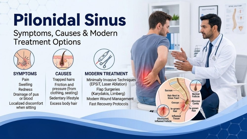
In plain language: A pilonidal sinus is a small tunnel beneath the skin near your tailbone that usually contains trapped hair. It is extremely common in young adults between 18 and 35 - particularly men who sit for long hours at a desk or in a car. It is not cancer, it is not sexually transmitted, and it is completely treatable. This page explains what causes it, how to spot it early, all available treatment options (including minimally invasive ones), and how to prevent it from coming back.
Quick Answers - Pilonidal Sinus
What Is a Pilonidal Sinus?
A pilonidal sinus is a small abnormal pocket or tunnel in the skin at the top of the buttock crease, an area surgeons call the natal cleft. The pocket typically contains hair follicles, dead skin cells and debris. Over time, this trapped material can become infected, forming an abscess (a painful, swollen, pus-filled lump) or a chronically draining sinus tract (a channel that intermittently leaks fluid or pus).
The condition was first described in detail during World War II, when thousands of army jeep drivers developed it from hours of bouncing on rough seats - earning it the nickname “jeep disease.” Today, the same problem affects students, software engineers, call-centre workers and anyone whose daily routine involves prolonged sitting on hard surfaces.
Who Gets Pilonidal Sinus?
Pilonidal disease overwhelmingly affects young adults between 18 and 35. After the age of 40, new cases become uncommon. Key risk factors include:
- Male sex - men are affected 3-4 times more often than women, largely because of coarser, denser body hair.
- Thick or stiff body hair - loose hair from the back and buttock area can be driven into the natal cleft skin by friction.
- Prolonged sitting - desk jobs, long commutes, or studying for hours increases pressure and sweating in the natal cleft.
- Obesity - deeper natal cleft, more moisture and greater friction combine to raise risk.
- Poor local hygiene - infrequent cleaning of the natal cleft area allows hair and debris to accumulate.
- Family history - a genetic tendency toward thick hair and deep natal cleft anatomy can run in families.
- Previous trauma - local injury or repeated microtrauma from sitting on hard surfaces can initiate the process.
Acute Abscess vs. Chronic Sinus - Two Presentations
Pilonidal disease shows up in two main ways, and understanding the difference matters because the treatment approach is different for each.
| Feature | Acute Pilonidal Abscess | Chronic Pilonidal Sinus |
|---|---|---|
| Onset | Sudden (hours to days) | Gradual (weeks to months) |
| Pain | Severe, throbbing, constant | Mild, intermittent, aching |
| Swelling | Tense, hot, red lump | Small pit(s) with surrounding thickening |
| Discharge | Pus if it ruptures | Intermittent clear or blood-tinged fluid |
| Fever | Often present | Usually absent |
| Sitting | Extremely difficult | Uncomfortable but manageable |
| Immediate treatment | Incision & drainage (I&D) | Planned definitive surgery |
Many patients first encounter pilonidal disease as an acute abscess. After the abscess is drained, about 40-50 % go on to develop a chronic sinus that keeps flaring up - which is when definitive surgical treatment becomes necessary.
Symptoms of Pilonidal Sinus
Early signs are easy to dismiss, especially in young people who may feel embarrassed to mention them. Watch for:
- A small dimple, pit or depression at the top of the buttock crease
- Recurring pain or tenderness near the tailbone, especially after sitting
- Swelling or a firm lump in the natal cleft that worsens over days
- Pus, blood or foul-smelling fluid draining from one or more openings
- Redness and warmth of the overlying skin
- Staining of undergarments with discharge
- Difficulty sitting comfortably in class, at work, or while driving
Symptoms typically flare during periods of prolonged sitting, hot weather (more sweating), or after local trauma such as a long bike ride.
How Is Pilonidal Sinus Diagnosed?
In most cases, a diagnosis is made by clinical examination alone. Your surgeon will look for characteristic midline pits, sinus openings, and surrounding induration (hardening) in the natal cleft. Additional investigations are rarely needed but may include:
- Ultrasound - to map the extent of an abscess cavity before drainage.
- MRI - for complex or recurrent disease where the surgeon needs to visualise deep tracts and rule out other conditions.
- Probe and methylene blue dye - used during surgery to trace the full extent of sinus tracts and ensure complete excision.
No blood test or biopsy is routinely required. The diagnosis is straightforward in experienced hands and can be confirmed within minutes of a clinical consultation.
Treatment Options for Pilonidal Sinus
1. Incision & Drainage (I&D) - For Acute Abscess
When a pilonidal abscess is tense and painful, the immediate priority is to release the pus. This is a quick bedside or OT procedure done under local anaesthesia. A small cut is made, pus is drained, and the wound is left open to heal. I&D provides rapid pain relief but does not cure the underlying sinus - about half of patients will eventually need a definitive procedure.
2. Wide Excision & Open Healing
The entire sinus tract and surrounding diseased tissue are cut out, and the wound is left open to heal by secondary intention (from the bottom up). This method has a low recurrence rate but recovery is slow - the open wound needs daily packing and dressing for 4-8 weeks. It remains a viable option for small, uncomplicated sinuses.
3. Limberg (Rhomboid) Flap
After excision of the sinus, a diamond-shaped skin flap from the adjacent buttock is rotated to cover the defect. The wound is closed primarily, which means faster healing (2-3 weeks) and a recurrence rate of only 3-5 %. The Limberg flap is one of the most widely studied and reliable techniques for moderate to severe pilonidal disease.
4. Karydakis Flap
An asymmetric, off-midline closure technique. The sinus is excised, and the wound edge is shifted away from the midline crease, which reduces friction, moisture and hair accumulation at the closure site. Recurrence rates are comparable to the Limberg flap (4-8 %), and the procedure flattens the natal cleft to discourage future hair entry.
5. Endoscopic Pilonidal Sinus Treatment (EPSiT)
A minimally invasive, camera-guided procedure. A small endoscope is inserted through the sinus opening, and the tract is cleaned, debrided and cauterised from within - without making a large external wound. Benefits include less pain, faster return to work (often within 3-5 days), and a small scar. Best suited for primary, non-complex sinuses.
6. Laser Ablation (SiLaC / FiLaC)
A radial laser fibre is inserted into the sinus tract and fires energy to destroy the tract lining from inside. Like EPSiT, this is a minimally invasive option with rapid recovery and a cosmetically small wound. It is particularly attractive for young patients concerned about scarring and downtime.
| Procedure | Wound | Recovery | Recurrence | Best For |
|---|---|---|---|---|
| I&D | Small open | 3-5 days | 40-50 % | Acute abscess (emergency) |
| Wide excision (open) | Large open | 4-8 weeks | 10-15 % | Small, primary sinus |
| Limberg flap | Closed | 2-3 weeks | 3-5 % | Moderate-severe disease |
| Karydakis flap | Closed | 2-3 weeks | 4-8 % | Recurrent disease, deep cleft |
| EPSiT | Minimal | 3-7 days | 8-15 % | Primary, non-complex sinus |
| Laser (SiLaC) | Minimal | 3-7 days | 10-18 % | Young patients, cosmetic concern |
How to Prevent Pilonidal Sinus from Coming Back
Regardless of which surgery you have, following these steps will significantly lower the chance of recurrence:
- Hair removal - regular shaving, depilatory cream or laser hair reduction in the natal cleft and lower back area. This is the single most important preventive measure.
- Keep the area clean and dry - daily washing with mild soap, thorough drying after bathing.
- Avoid prolonged sitting - take a 5-minute standing break every 45-60 minutes during work or study sessions.
- Maintain a healthy weight - reducing body weight decreases cleft depth and local sweating.
- Wear loose, breathable clothing - tight jeans and synthetic underwear trap heat and moisture.
- Complete all follow-up visits - your surgeon needs to confirm full healing and check for early recurrence.
Red Flags - See a Doctor Urgently
- Rapidly enlarging, tense swelling near the tailbone with fever (> 100.4 °F / 38 °C)
- Spreading redness across the buttock or lower back (cellulitis)
- Inability to sit, walk or lie down comfortably
- Foul-smelling discharge that does not improve after 48 hours of antibiotics
- Recurrent abscesses (3 or more episodes) - indicates a chronic sinus needing definitive surgery
Reassuring Signs
- A tiny, painless midline pit with no discharge - may be monitored with hygiene measures
- Mild tenderness that settles with warm compresses and improved hygiene
- Post-surgical wound healing steadily with decreasing discharge
- No fever, no spreading redness, no systemic illness
What Happens If Pilonidal Sinus Is Ignored?
- Recurrent abscesses - the infection returns every few weeks or months, each time more painful than before.
- Expanding sinus network - a single tract can branch into multiple tracts with several external openings, making eventual surgery larger and more complex.
- Chronic draining wound - persistent discharge stains clothing, causes odour and affects social confidence - a real concern for college students and young professionals.
- Scarring and skin changes - repeated infections damage the skin, making flap repairs more difficult later on.
- Lost productivity - missed college days, sick leave from work, inability to sit through exams or meetings.
- Extremely rare: malignant change - decades of untreated chronic pilonidal sinus has been associated, in very rare case reports, with squamous cell carcinoma. This is not a reason to panic but is a reason not to ignore the condition indefinitely.
Pilonidal Sinus in India - Why It Matters Here
India has one of the world’s largest populations of young adults in sedentary desk jobs. The IT sector alone employs over 5 million professionals who sit 8-10 hours a day in air-conditioned offices. Combine this with a genetically hairy phenotype common among South Asian men, and you have a recipe for pilonidal disease.
Yet many young Indians delay treatment for months or even years because of:
- Embarrassment - the location makes it awkward to discuss, especially with a GP of the opposite gender.
- Misdiagnosis - the swelling is often mistaken for a “boil” and treated with repeated courses of antibiotics without addressing the underlying sinus.
- Fear of surgery - outdated information about large open wounds and weeks of bed rest discourages patients from seeking definitive care.
- Lack of awareness - most people have never heard of pilonidal sinus until they develop one themselves.
The reality is that modern minimally invasive options like EPSiT and laser ablation allow most patients to return to their desk within a week. Early consultation with an experienced surgeon makes all the difference.
Pilonidal Sinus Treatment in Vadodara
Dr Samir Contractor offers the full range of pilonidal sinus treatments at Sterling Hospital, Vadodara - from emergency abscess drainage to flap repairs and minimally invasive procedures.
- Experienced in all standard and advanced techniques: wide excision, Limberg flap, Karydakis flap, EPSiT, and laser ablation
- Day-care and short-stay options available - most patients go home the same day or within 24 hours
- Comprehensive post-operative follow-up with wound-care guidance and recurrence-prevention counselling
- Insurance and cashless facility available at Sterling Hospital for eligible patients
- Patients from Anand, Bharuch, Godhra, Dahod and surrounding districts regularly visit for specialised anorectal care
ગુજરાતીમાં સામાન્ય પ્રશ્નો (FAQs in Gujarati)
પાઇલોનાઇડલ સાઇનસ એ કમરના નીચેના ભાગમાં (પૂંછડીના હાડકા પાસે) ત્વચાની નીચે બનતી એક નાની ટનલ છે જેમાં વાળ ફસાય છે. “પાઇલોનાઇડલ”નો અર્થ છે “વાળનો માળો.” આ ચેપ લાગવાથી દુખાવો, સોજો અને પરુ નીકળી શકે છે.
18 થી 35 વર્ષના યુવાન પુરુષોમાં આ સૌથી સામાન્ય છે. IT પ્રોફેશનલ્સ, વિદ્યાર્થીઓ અને ડ્રાઇવરો જેવા લાંબો સમય બેસતા લોકોમાં, તેમજ વધુ વાળ ધરાવતા અને મેદસ્વી વ્યક્તિઓમાં જોખમ વધારે છે.
ખૂબ હળવા કિસ્સામાં એન્ટીબાયોટિક્સ અને સ્વચ્છતાથી રાહત મળી શકે. પરંતુ એક વાર ફોલ્લી (abscess) અથવા સાઇનસ ટ્રેક્ટ બની ગયા પછી, કાયમી ઈલાજ માટે સર્જરી જરૂરી છે.
ફ્લૅપ સર્જરી (Limberg/Karydakis) પછી ફરી થવાની શક્યતા 3-8 % છે. નિયમિત વાળ દૂર કરવા, સ્વચ્છતા જાળવવી અને વજન નિયંત્રણ રાખવાથી આ જોખમ વધુ ઘટાડી શકાય છે.
ડૉ. સમીર કોન્ટ્રાક્ટર સ્ટર્લિંગ હોસ્પિટલ, વડોદરામાં પાઇલોનાઇડલ સાઇનસની તમામ આધુનિક સારવાર આપે છે - ફ્લૅપ સર્જરી, EPSiT અને લેસર સહિત. મોટાભાગના દર્દીઓ તે જ દિવસે ઘરે જઈ શકે છે.
મિનિમલી ઇન્વેસિવ પ્રક્રિયા (EPSiT, લેસર) પછી 5-7 દિવસમાં ડેસ્ક જોબ પર પાછા ફરી શકાય. ફ્લૅપ સર્જરી પછી 2-3 અઠવાડિયા મર્યાદિત બેસવું જરૂરી છે. ડૉક્ટર તમારી પરિસ્થિતિ પ્રમાણે માર્ગદર્શન આપશે.
Dealing with a Painful Lump Near Your Tailbone?
Early treatment means a smaller wound, faster recovery and lower recurrence. Talk to Dr Samir Contractor today.