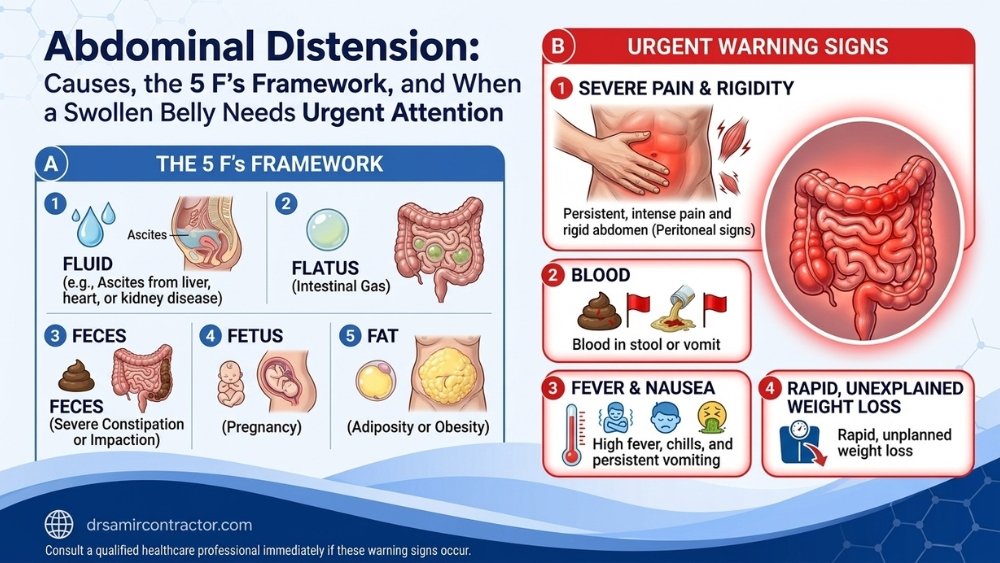
In simple terms: If your belly is visibly bigger than normal - not just a feeling of fullness, but something you or others can actually see - that is abdominal distension. It can happen slowly over weeks or suddenly within hours. Doctors use a classic checklist called the 5 F's (Fat, Fluid, Flatus, Faeces, Fetus) to narrow down the cause. While some causes are harmless, others - like fluid accumulation from liver disease, bowel obstruction, or an abdominal mass - require urgent investigation. This page explains how to tell the difference and when you should not wait.
✦ Quick Answers
What Exactly Is Abdominal Distension?
Abdominal distension refers to a visible, measurable enlargement of the abdomen. It is not a diagnosis in itself but rather a physical sign that something has changed inside the abdominal cavity or the abdominal wall. When a patient or their family member says "the stomach has become big" or "the belly looks swollen," they are describing distension.
The key distinction from abdominal bloating is objectivity. Bloating is a subjective sensation - you feel full, tight, or pressurised even though your waistline may not have changed. Distension, on the other hand, is something that can be seen and measured. Your trousers become tighter. A tape measure around the navel shows an increase. In clinical practice, this distinction matters enormously because the list of causes - and the urgency of investigation - differs significantly.
Distension may develop gradually over weeks to months (as in chronic liver disease or a slow-growing ovarian cyst) or appear suddenly within hours (as in acute bowel obstruction or a ruptured organ). The speed of onset often provides the first crucial diagnostic clue.
It is important to understand that abdominal distension is not a diagnosis - it is a clinical sign that demands a systematic search for the underlying cause. The abdomen is a large cavity containing the stomach, intestines, liver, spleen, kidneys, pancreas, uterus (in women), major blood vessels, and the peritoneal membrane that lines everything. Any pathology affecting these structures can manifest as visible abdominal swelling. This is why a careful, structured clinical approach is essential rather than dismissing the symptom as "just gas" or "weight gain."
In clinical practice, we measure abdominal distension by recording the abdominal girth at the level of the umbilicus (navel) with a flexible tape measure. Serial measurements taken on different days or weeks help determine whether the distension is stable, fluctuating, or progressively increasing - a critical distinction that guides the urgency of investigation.
Distension vs Bloating - Why the Difference Matters
Patients frequently use "bloating" and "distension" interchangeably, but clinically they represent different problems with different implications:
- Bloating (subjective): Often functional, linked to irritable bowel syndrome (IBS), food intolerances, aerophagia, or dysmotility. Typically fluctuates during the day and improves overnight. Rarely signals serious organic disease when present in isolation.
- Distension (objective): Always warrants investigation, especially when progressive. May indicate fluid accumulation, obstruction, mass, or organ enlargement. Demands imaging, blood work, and sometimes invasive assessment.
If you are experiencing a feeling of fullness without visible enlargement, our detailed guide on abdominal bloating may be more relevant. This page focuses on the causes, evaluation, and treatment of true, measurable abdominal swelling.
The 5 F's Framework - A Systematic Approach to a Distended Abdomen
Medical students worldwide learn the "5 F's" mnemonic as a structured way to think through the differential diagnosis of abdominal distension. Each "F" represents a broad category of causes:
| The "F" | What It Means | Common Examples | Onset |
|---|---|---|---|
| Fat | Central (visceral) obesity or lipodystrophy | Metabolic syndrome, Cushing's syndrome, truncal obesity | Gradual (months-years) |
| Fluid | Free fluid in the peritoneal cavity (ascites) | Cirrhosis, heart failure, ovarian cancer, TB peritonitis, nephrotic syndrome | Gradual to subacute |
| Flatus | Trapped gas within the bowel lumen | Bowel obstruction, pseudo-obstruction (Ogilvie's), severe dysmotility, aerophagia | Hours to days |
| Faeces | Accumulated stool in the colon | Chronic constipation, faecal impaction, megacolon, Hirschsprung's disease (in children) | Days to weeks |
| Fetus | Pregnancy or uterine enlargement | Normal pregnancy, large fibroids, molar pregnancy | Gradual |
| Fatal (6th F) | Mass or tumour | Ovarian mass, colorectal tumour, mesenteric cyst, retroperitoneal sarcoma, massive hepatosplenomegaly | Gradual to subacute |
This framework is not just academic - it directly guides the clinical examination and choice of investigations. For example, a surgeon will check for shifting dullness (fluid), tympanic percussion (gas), or a palpable mass (tumour) during the physical exam, and each finding points toward a specific "F."
In my practice at Sterling Hospital, I routinely use this framework during the initial assessment. Within minutes of examining a patient with a distended abdomen, the 5 F's approach allows me to narrow the possibilities and order targeted investigations rather than a scattergun battery of tests. Patients benefit from faster diagnosis and reduced unnecessary testing.
It is worth noting that more than one "F" can be present simultaneously. A patient with chronic liver disease may have both fluid (ascites) and flatus (from gut dysmotility associated with portal hypertension). An elderly patient may have fat, faeces (chronic constipation), and an undiagnosed mass all contributing to their distended abdomen. This overlap underscores the importance of thorough evaluation rather than stopping at the first explanation.
Major Causes of Abdominal Distension - Explained by Mechanism
1. Ascites (Fluid Accumulation)
Ascites is the pathological accumulation of free fluid in the peritoneal cavity. It is one of the most important causes of progressive abdominal distension and always demands thorough investigation. The most common cause in India is chronic liver disease (cirrhosis), often related to alcohol use or viral hepatitis B/C. Other causes include:
- Congestive heart failure (right-sided or biventricular)
- Ovarian malignancy - often presents as "abdominal fullness" before other symptoms appear
- Tuberculous peritonitis - particularly relevant in the Indian population
- Nephrotic syndrome and severe hypoalbuminaemia
- Peritoneal carcinomatosis from gastrointestinal cancers
Clinical signs of ascites include a fluid thrill, shifting dullness on percussion, and an everted umbilicus in tense ascites. A diagnostic tap (paracentesis) can determine whether the fluid is a transudate or exudate, guiding further management.
The volume of ascites can range from a few hundred millilitres (detectable only by ultrasound) to over 10-15 litres in tense ascites. Patients with tense ascites often describe difficulty breathing when lying flat (orthopnoea), reduced oral intake because the stomach is compressed, and significant discomfort from the sheer weight and pressure of the fluid. Therapeutic large-volume paracentesis - draining several litres under sterile conditions - provides immediate relief and is both diagnostic and therapeutic.
In India, it is crucial to include tuberculous peritonitis in the differential for any young patient presenting with ascites, low-grade fever, and weight loss. The ascitic fluid in TB typically shows high protein, high lymphocyte count, and elevated adenosine deaminase (ADA) levels. Peritoneal biopsy may be needed for confirmation in doubtful cases.
2. Bowel Obstruction (Gas and Fluid Trapped Within the Gut)
Mechanical bowel obstruction produces distension because gas and intestinal contents cannot move forward. The abdomen becomes tympanic (drum-like) on percussion. This is a surgical emergency that may require urgent intervention.
- Small bowel obstruction: Most commonly caused by adhesions from previous surgery, hernias, or rarely, gallstone ileus. Symptoms include colicky pain, vomiting (often bilious), and absolute constipation. Read more: Small-Bowel Obstruction Warning.
- Large bowel obstruction: Colorectal cancer is the most common cause in adults over 50. Other causes include volvulus (sigmoid or caecal), diverticular stricture, and faecal impaction. See: Bowel Obstruction Warning Signs.
Any patient with distension combined with absolute constipation (no passage of gas or stool), vomiting, and colicky pain needs immediate hospital evaluation - delays can lead to bowel strangulation, ischaemia, and perforation.
3. Abdominal Masses and Organomegaly
A palpable mass can cause visible asymmetric or generalised distension. Common examples include:
- Large ovarian cysts or tumours (can grow to enormous size before causing symptoms)
- Massive hepatomegaly (enlarged liver - from cancer, fatty liver, or congestive hepatopathy)
- Splenomegaly (chronic myeloid leukaemia, portal hypertension, kala-azar)
- Mesenteric cysts, retroperitoneal tumours, or pseudomyxoma peritonei
A particularly important clinical scenario in Indian women is the large ovarian cyst or tumour. Because these masses grow slowly within the pelvis and then extend into the abdomen, patients often attribute the gradual increase in abdominal size to weight gain. It is not uncommon to encounter ovarian masses weighing 5-15 kg by the time the patient seeks medical attention. Cross-sectional imaging (CT or MRI) is essential to characterise these masses before planning surgery.
Massive hepatomegaly and splenomegaly deserve special mention. In parts of India where malaria, kala-azar (visceral leishmaniasis), and chronic myeloproliferative disorders are prevalent, the liver or spleen can enlarge to the point where it occupies much of the abdominal cavity. Palpation during physical examination can identify these enlarged organs, and ultrasound confirms the diagnosis.
4. Severe Constipation and Faecal Loading
Chronic constipation can cause enough stool to accumulate in the colon that the abdomen becomes visibly distended. This is especially common in elderly patients, those on opioid medications, and individuals with a low-fibre diet. In extreme cases, faecal impaction requires manual disimpaction or surgical intervention. For more on related symptoms, see recurrent abdominal discomfort.
5. Central Obesity and Fat Distribution
While obesity is the most common cause of a "big belly" overall, it is usually apparent and chronic. However, rapid central weight gain - especially with purple striae, moon face, and buffalo hump - may indicate Cushing's syndrome, which requires hormonal evaluation.
6. Functional Abdominal Distension
Some patients exhibit measurable abdominal distension without any identifiable organic cause. This is classified as functional bloating or functional distension under Rome IV criteria. It is a diagnosis of exclusion - meaning all serious causes must be ruled out first. The mechanism may involve diaphragmatic-pelvic dyssynergia, where the diaphragm descends and the anterior abdominal wall relaxes paradoxically.
Red Flags - When Abdominal Distension Requires Urgent Medical Attention
- Sudden onset with severe abdominal pain - may indicate obstruction, perforation, or vascular catastrophe
- Absolute constipation (no gas, no stool) - hallmark of complete bowel obstruction
- Persistent vomiting, especially if bilious (green) or faeculent (foul-smelling)
- Rapid progression over hours to days with tense abdomen
- Jaundice (yellow skin/eyes) + distension - suggests liver disease with ascites
- Unintentional weight loss combined with increasing abdominal girth - malignancy must be excluded
- Blood in stool or vomit alongside distension
- Fever with distension - peritonitis, infected ascites, or intra-abdominal abscess
- Inability to lie flat due to abdominal tightness and breathlessness
- New-onset leg swelling combined with abdominal distension - may indicate IVC compression, liver disease, or pelvic mass
Any of these combinations requires same-day medical evaluation. Do not adopt a "wait and see" approach.
When to Worry vs When to Wait - A Comparison
| Feature | Likely Benign / Functional | Potentially Serious / Organic |
|---|---|---|
| Onset | Fluctuates through the day; better in morning | Constant or progressively worsening |
| Duration | Comes and goes for months/years | New symptom, weeks to months, worsening |
| Associated symptoms | Mild discomfort, excess gas, normal appetite | Pain, vomiting, weight loss, jaundice, blood in stool |
| Bowel habit | Irregular but gas and stool still pass | Complete cessation of gas and stool |
| Physical exam | Soft abdomen, no masses, no fluid signs | Tense abdomen, shifting dullness, palpable mass, tenderness |
| Weight change | Stable weight | Unexplained weight loss despite larger abdomen |
Reassuring Signs - When Distension Is Likely Not Dangerous
- Belly is flat in the morning and progressively distends by evening, then resets overnight - this pattern strongly suggests functional distension or dietary-related gas
- Distension improves after passing gas or having a bowel movement
- No associated pain, vomiting, weight loss, or change in stool colour
- You are otherwise well with normal appetite and energy
- Longstanding pattern (years) without progression
Even if your symptoms seem benign, a baseline evaluation by a specialist is worthwhile to rule out treatable conditions. Many patients find significant relief once the right cause is identified.
How Is Abdominal Distension Diagnosed?
Diagnosis follows a structured approach:
- History: Onset (sudden vs gradual), duration, associated symptoms (pain, vomiting, constipation, weight change, jaundice), dietary habits, alcohol intake, medication use, surgical history, menstrual history in women.
- Physical examination: Inspection (shape, symmetry, visible peristalsis), palpation (tenderness, masses, organ enlargement), percussion (tympany vs dullness, shifting dullness for fluid), auscultation (bowel sounds - absent in ileus, high-pitched/tinkling in obstruction).
- Blood investigations: Complete blood count, liver function tests, serum albumin, kidney function, thyroid profile, tumour markers (CA-125, CEA) when malignancy is suspected.
- Imaging:
- Abdominal ultrasound - first-line investigation; detects free fluid, organomegaly, masses, and gallstones
- Plain abdominal X-ray - shows dilated loops (obstruction), air-fluid levels, and free gas under diaphragm (perforation)
- CT scan of abdomen - gold standard for detailed evaluation of masses, obstruction site, and peritoneal disease
- Endoscopic evaluation: Colonoscopy to evaluate large bowel causes (cancer, strictures, volvulus). Upper GI endoscopy when gastric outlet obstruction or upper GI pathology is suspected.
- Ascitic fluid analysis: If fluid is detected, paracentesis determines the cause - the serum-ascites albumin gradient (SAAG) differentiates portal hypertension-related ascites from other causes.
Treatment of Abdominal Distension - Addressing the Underlying Cause
There is no single treatment for distension because it is a sign, not a disease. Effective treatment requires identifying and managing the root cause:
- Ascites: Sodium restriction, diuretics (spironolactone, furosemide), therapeutic paracentesis for tense ascites, TIPS procedure for refractory cases, and treatment of the underlying liver/heart/kidney disease.
- Bowel obstruction: Initial management with nasogastric decompression, IV fluids, and nil by mouth. Surgical intervention - including colorectal surgery - is required when conservative measures fail or when strangulation is suspected.
- Masses/tumours: Surgical excision (often laparoscopic), followed by appropriate oncological treatment if malignant.
- Severe constipation: Osmotic laxatives, enemas, dietary fibre supplementation, adequate hydration. Manual disimpaction in severe faecal loading.
- Functional distension: Biofeedback therapy for abdomino-phrenic dyssynergia, low-FODMAP diet trial, prokinetic agents, and management of underlying IBS.
- Central obesity: Structured weight management, dietary modification, exercise, and metabolic assessment.
What Happens If Abdominal Distension Is Ignored?
The consequences of ignoring distension depend on the cause, but in several scenarios, delay is dangerous:
- Untreated bowel obstruction can progress to bowel strangulation, ischaemia, necrosis, and perforation - a life-threatening emergency requiring emergency surgery with higher mortality.
- Undiagnosed ascites may represent advanced liver disease (where early intervention can slow progression) or occult malignancy (where delayed diagnosis reduces treatment options).
- Growing masses become harder to remove surgically and may become inoperable if caught late.
- Chronic severe constipation can lead to stercoral ulceration, perforation, and megacolon - all avoidable with timely management.
The take-home message: visible, progressive abdominal swelling is not something to dismiss. Early evaluation saves lives, reduces the need for emergency surgery, and often leads to simpler, less invasive treatment.
Abdominal Distension in Special Populations
Elderly Patients
In patients over 65, new-onset abdominal distension deserves particularly careful evaluation. The threshold for suspecting malignancy should be low, especially with concurrent weight loss or change in bowel habit. Sigmoid volvulus - a condition where the sigmoid colon twists on itself - is more common in elderly, institutionalised, or neurologically impaired patients and presents with dramatic, rapid-onset distension. Ogilvie's syndrome (acute colonic pseudo-obstruction) is another cause seen in hospitalised elderly patients, producing massive colonic dilatation without a mechanical blockage.
Women of Reproductive Age
In any woman of reproductive age presenting with abdominal distension, pregnancy must be excluded first - this is a fundamental clinical principle. Beyond pregnancy, ovarian pathology (cysts, tumours, endometriomas) is a leading cause of lower abdominal distension in women. Ovarian cancer is sometimes called the "silent killer" because its early symptoms - mild distension, vague pelvic discomfort, early satiety - are often attributed to less serious conditions. A high index of suspicion combined with pelvic ultrasound and CA-125 levels can aid early detection.
Post-Surgical Patients
Patients with a history of abdominal surgery are at risk for adhesive bowel obstruction, which can present months or even years after the original operation. Adhesions are the most common cause of small bowel obstruction worldwide. Any patient who has had previous abdominal surgery and develops distension with colicky pain and vomiting should be evaluated urgently. A CT scan with oral contrast can determine the site and severity of the obstruction and guide the decision between conservative management and reoperation.
Concerned about abdominal distension? Dr Samir Contractor provides thorough clinical evaluation with same-week appointments at Sterling Hospital, Vadodara.
Abdominal Distension in the Indian Context
Several factors make the evaluation of abdominal distension particularly important in India:
- Tuberculosis: TB peritonitis remains a significant cause of ascites in India, especially in younger patients. Unlike in Western countries where liver cirrhosis dominates, TB must always be considered in the differential diagnosis of unexplained ascites with fever and weight loss.
- Hepatitis B & C burden: India has a substantial population of chronic hepatitis carriers. Many patients present with ascites as the first sign of previously undiagnosed cirrhosis.
- Alcohol-related liver disease: Rising alcohol consumption, particularly among men aged 30-55, has led to increasing rates of alcoholic cirrhosis presenting with ascites and distension.
- Late presentation of cancers: Ovarian, gastric, and colorectal cancers in India are frequently diagnosed at advanced stages, often because early symptoms like mild distension were attributed to "gas" or "acidity."
- Dietary factors: High-carbohydrate, legume-heavy vegetarian diets can produce significant intestinal gas, leading to functional distension. While this is generally benign, it often coexists with and masks more serious pathology.
- Self-medication culture: Prolonged use of over-the-counter antacids, laxatives, and Ayurvedic preparations can mask progressive distension until the underlying disease is advanced.
- Tropical infections: Amoebic liver abscess, hydatid cyst, and visceral leishmaniasis (kala-azar) can cause hepatosplenomegaly and distension - conditions less common in Western practice.
Common Questions in Gujarati & Hinglish
If your belly is getting progressively larger, do not ignore it. It could be fluid, gas, or a growth. Get an ultrasound done and consult a surgeon. Early detection makes treatment much simpler.
Water in the belly (ascites) usually means the liver, heart, or kidneys are not working properly. Sometimes it can be from TB or cancer. A simple test of the fluid tells the doctor the exact cause.
If your tummy looks visibly swollen - not just a feeling - it could be more than gas. Gas-related distension usually comes and goes, but if it stays or gets worse, you need a check-up to rule out obstruction or fluid.
Severe constipation can fill the colon with so much stool that the belly becomes visibly big. But if you are passing stool normally and the belly is still growing, the cause is something else - fluid, a mass, or organ enlargement.
Yes, when the liver is damaged (cirrhosis), fluid collects in the belly. This is called ascites. The belly becomes tight, the navel may pop out, and you may feel breathless. Medicines and sometimes draining the fluid can help.
Sudden swelling of the belly is a warning sign. It can mean bowel obstruction, internal bleeding, or a burst organ. Go to the hospital immediately - do not wait at home or try home remedies.
Frequently Asked Questions
Bloating is a subjective feeling of abdominal fullness or pressure - the patient feels uncomfortable, but the belly may not look larger. Distension is an objective, visible enlargement of the abdomen that can be measured. Distension carries a broader differential diagnosis and typically warrants more urgent investigation.
Using the 5 F's framework: Fat (obesity), Fluid (ascites from liver disease, heart failure, or cancer), Flatus (trapped intestinal gas from obstruction or dysmotility), Faeces (severe constipation or impaction), and Fetus (pregnancy). The sixth "F" is Fatal - meaning tumours or abdominal masses.
Yes. Significant gas accumulation - from aerophagia, small intestinal bacterial overgrowth (SIBO), or bowel dysmotility - can produce measurable distension. However, when gas causes severe or sudden distension with pain and inability to pass flatus, bowel obstruction must be ruled out. See also: Gas and Flatulence.
In small bowel obstruction, distension typically develops over hours to 1-2 days. In large bowel obstruction, it may build up over several days. The speed depends on the level and completeness of the obstruction.
Ascites causes a diffuse, heavy fullness. The abdomen feels tight (especially in tense ascites), and patients often report difficulty lying flat, shortness of breath, and a "sloshing" sensation when moving. The flanks appear full and bulging when lying on the back.
No. Most cases of abdominal distension are caused by benign conditions such as constipation, gas, obesity, or functional distension. However, persistent, progressive distension combined with weight loss, loss of appetite, or new-onset ascites should prompt investigation to exclude malignancy.
Yes, if distension is accompanied by severe pain, vomiting, inability to pass gas or stool, fever, blood in stool/vomit, or rapid worsening over hours. These symptoms suggest possible bowel obstruction, perforation, or peritonitis - all surgical emergencies.
Yes. Post-operative distension can result from ileus (temporary paralysis of bowel function), adhesive obstruction, or intra-abdominal fluid collection. Some post-operative distension is expected and resolves within 2-4 days, but persistent or worsening distension requires evaluation.
During physical examination, the doctor checks for shifting dullness (dull sound shifts when you roll to one side) and a fluid thrill (a ripple felt across the abdomen). Ultrasound can detect as little as 100-200 mL of free fluid and is the most reliable bedside test.
Common tests include complete blood count, liver function tests (AST, ALT, bilirubin), serum albumin and protein, kidney function (creatinine, BUN), thyroid profile, and tumour markers (CA-125 for ovarian pathology, CEA for colorectal cancer) when malignancy is suspected.
Stress can worsen functional distension through its effects on gut motility, visceral hypersensitivity, and breathing patterns (stress-related aerophagia). However, stress alone does not cause the serious organic causes of distension. If distension is new or progressive, it should be investigated regardless of stress levels.
Many women experience some degree of abdominal distension premenstrually due to hormonal fluid retention and altered bowel motility. This is usually mild, cyclical, and self-resolving. Severe or persistent distension around periods may warrant evaluation for endometriosis or ovarian pathology.
If distension is caused by gas or functional bloating, dietary modification (reducing high-FODMAP foods, carbonated drinks, and poorly digested carbohydrates) can help significantly. However, dietary changes will not address distension caused by ascites, obstruction, or masses - these require medical or surgical treatment.
A CT scan of the abdomen and pelvis is the gold standard for evaluating unexplained distension. It can identify the site and cause of obstruction, detect masses and their extent, quantify ascites, and reveal peritoneal disease. Contrast-enhanced CT provides the most information.
Yes. In infants, distension may indicate Hirschsprung's disease, necrotising enterocolitis, or intestinal atresia. In older children, it can result from coeliac disease, lactose intolerance, worm infestation (common in India), or rarely, abdominal tumours such as Wilms' tumour or neuroblastoma.
If distension is new, persistent (lasting more than 2 weeks), progressive, or accompanied by any red-flag symptoms (pain, vomiting, constipation, weight loss, jaundice), consult a doctor within days, not weeks. Sudden distension with pain is an emergency requiring same-day evaluation.
Yes, a strong one. Chronic liver disease (cirrhosis) is one of the most common causes of ascites, which presents as progressive abdominal distension. The liver's inability to produce albumin and the elevated portal venous pressure both contribute to fluid accumulation in the abdomen.
Recurrence depends on the underlying cause. Ascites from cirrhosis often recurs unless the liver disease is controlled. Obstruction from adhesions can recur. Functional distension may wax and wane. Post-surgical removal of a mass or tumour usually resolves distension permanently if the disease does not recur.
Related Pages
Do not ignore a swollen belly. Schedule a clinical evaluation with Dr Samir Contractor today.
Medical Disclaimer: The information on this page is provided for educational and informational purposes only. It is not a substitute for professional medical advice, diagnosis, or treatment. Always seek the advice of a qualified healthcare provider with any questions you may have regarding a medical condition. Never disregard professional medical advice or delay seeking it because of something you have read on this website. If you think you may have a medical emergency, call your doctor or local emergency services immediately.
© 2026 Dr Samir Contractor. All rights reserved.