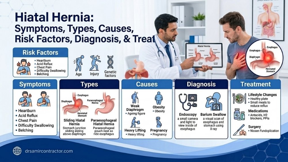
A hiatal hernia occurs when part of the stomach pushes through an opening in the diaphragm (hiatus) into the chest cavity. The most common type is closely linked to GERD and heartburn. Most small hiatal hernias do not require surgery. Large or symptomatic hernias - particularly those causing severe reflux or at risk of gastric strangulation - need surgical repair.
✦ Quick Answers
The diaphragm is the dome-shaped muscle separating the chest from the abdomen. It has a natural opening called the hiatus through which the esophagus passes before connecting to the stomach. A hiatal hernia occurs when the stomach or part of it pushes through this opening into the chest.
Hiatal hernias are among the most common anatomical findings on upper GI endoscopy and radiology. Many are incidental - found during investigation of heartburn or other symptoms. Understanding the type of hernia determines whether medical or surgical management is appropriate.
The Four Types of Hiatal Hernia
Type I - Sliding (Most Common)
- 95% of all hiatal hernias
- Esophagogastric junction and part of stomach slide into chest
- Disrupts the anti-reflux mechanism
- Most common cause of GERD
- Usually managed medically
- Surgery for symptomatic refractory cases
Type II - Paraesophageal (Rolling)
- GE junction stays in normal position
- Gastric fundus herniates beside esophagus
- Less common but higher risk
- Can cause intermittent obstruction
- Risk of gastric volvulus (twisting)
- Surgery usually recommended
Type III - Mixed
- Combination of sliding and paraesophageal
- Both GE junction and fundus herniate
- Significant reflux and obstruction symptoms
- Higher complication risk
- Surgical repair recommended
Type IV - Complex
- Other abdominal organs herniate (colon, spleen)
- Rare but most severe
- May cause significant respiratory symptoms
- Chest heaviness, shortness of breath
- Surgical repair required
Symptoms of Hiatal Hernia
Small sliding hernia (Type I)
- Often asymptomatic - found incidentally during endoscopy
- Heartburn and regurgitation - from disrupted anti-reflux mechanism
- Post-meal fullness
- Symptoms worsened by bending, lying flat, and large meals
Large hernia (Types II-IV)
- Chest pressure or discomfort after eating
- Difficulty swallowing - from mechanical compression
- Shortness of breath - when hernia compresses the lungs
- Vomiting or retching that does not produce vomit (obstructive)
- Severe upper abdominal or chest pain - suggests volvulus (surgical emergency)
- Anaemia from chronic bleeding from the herniated stomach lining (Cameron lesions)
What Causes Hiatal Hernia?
- Age-related weakening of the hiatal musculature - the most common cause; prevalence increases markedly above age 50
- Increased intra-abdominal pressure - from obesity, chronic cough, straining (constipation), heavy lifting, and pregnancy
- Congenital weakness - some patients are born with a larger hiatal opening
- Previous esophageal or gastric surgery - can disturb the normal hiatal anatomy
- Trauma - rare cause; blunt abdominal trauma can force stomach into chest
Red Flags - When Hiatal Hernia Needs Urgent Attention
Seek emergency care if a known hiatal hernia causes:
- Sudden severe chest or upper abdominal pain - possible volvulus
- Inability to swallow or vomit (retching without vomiting) - obstructed hernia
- Shortness of breath with chest pain from a large hernia
- Signs of gastric strangulation - acute onset, severe pain, collapse
A paraesophageal hernia with acute onset severe symptoms is a surgical emergency. Early diagnosis and repair prevents catastrophic complications including gastric necrosis.
How Is Hiatal Hernia Diagnosed?
- Upper GI Endoscopy - identifies the hernia, measures its size, assesses the gastric mucosa for Cameron lesions, and evaluates GERD complications (esophagitis, Barrett's)
- Barium swallow - shows the anatomy of the hernia, its size, and the esophagogastric junction relationship; identifies sliding and paraesophageal hernias clearly
- CT scan of chest and abdomen - essential for large or complex hernias; shows which organs are herniated and guides surgical planning
- Esophageal manometry - assesses LES pressure and esophageal motility before surgical repair
Treatment of Hiatal Hernia
Medical management (Type I, asymptomatic or mild)
- PPI therapy for GERD symptoms - 30-60 minutes before breakfast
- Lifestyle modification: smaller meals, no eating 2-3 hours before sleep, elevate head of bed, weight loss
- Alginate antacids for regurgitation specifically
- H. pylori testing and eradication if positive
- Regular follow-up and endoscopic surveillance for Barrett's if present
Surgical management - Laparoscopic Hiatal Hernia Repair + Fundoplication
Surgery is performed through 4-5 small laparoscopic incisions. The herniated stomach is reduced from the chest back into the abdomen. The enlarged hiatal opening is repaired (cruroplasty - suturing the diaphragmatic crura together). A partial or full fundoplication (stomach wrap) is added to reinforce the anti-reflux mechanism.
Indications for surgery:
- Symptomatic paraesophageal hernia (Types II-IV) - due to volvulus risk
- Large sliding hernia with refractory GERD not responding to PPI therapy
- Confirmed GERD with significant hiatal hernia and patient preference for surgical correction
- Cameron lesions causing chronic anaemia from hernia-related bleeding
- Respiratory symptoms from large hernia compressing the lung
Surgical outcomes: Laparoscopic repair has an excellent safety profile. Hospital stay is typically 1-3 days. Full recovery in 2-4 weeks. Long-term relief of GERD symptoms and hernia-related symptoms is achieved in the majority of patients.
What Happens If a Hiatal Hernia Is Left Untreated?
- Worsening GERD - esophagitis, stricture, Barrett's esophagus from ongoing acid exposure
- Cameron lesions - erosions at the hernial neck causing chronic blood loss and iron deficiency anaemia
- Gastric volvulus - acute twisting of the herniated stomach; a life-threatening emergency requiring urgent surgery
- Progressive respiratory compromise - for large hernias, continued lung compression
Frequently Asked Questions
Hiatal Hernia in India
India-specific context
- Hiatal hernia is frequently missed in Indian patients because they are managed for years with antacids without an endoscopy that would identify the structural cause of their GERD
- The increasing prevalence of obesity in urban India - particularly central obesity - is a major driver of new hiatal hernia development and worsening of existing hernias
- Cameron lesion anaemia from large hiatal hernias is under-recognised - unexplained anaemia in older Indian patients warrants upper GI endoscopy
- Large paraesophageal hernias presenting as acute chest pain and inability to vomit are occasionally misdiagnosed as cardiac events - the history and endoscopy/CT distinguish them
Seek Care in Vadodara
Hiatal hernia identified on endoscopy or CT - consult Dr Samir Contractor at Sterling Hospital, Vadodara for surgical assessment and laparoscopic repair when indicated.
Desi Patient Questions
Ha - stomach noo ek bhag diaphragm (parda) thi upar chest cavity ma jay jaay chhe. Ee anti-reflux valve nu position change kare chhe ane GERD produce kare chhe. Majority cases (Type I) medically manage thay chhe. Large or paraesophageal type surgery mate recommend thay chhe.
Necessarily nahi. Small Type I hernia - PPI + lifestyle thi manage thay chhe. Operation jyaré: hernia motu hoy, symptoms medicine thi control na thay, ya paraesophageal type hoy (volvulus risk). Doctor proper assessment karshe - endoscopy + barium swallow + symptoms basis par recommendation milshe.
Laparoscopic surgery chhe - 4-5 small holes (5-10mm) thi thay chhe. Stomach pachi abomen ma lakho, diaphragm repair karyo, ane fundoplication karo to reflux stop thay. 1-3 days hospital. 2-4 weeks ma normal khavanu. Minimally invasive - scars minimal.